Information for patients

Patient resources

Tests, findings
and next steps.

I explain what I look for, how I interpret a result and what usually happens next in New Zealand. Explore a diagram or keep a short guide for later.

Browse the library
Your saved guidesNo saved favourites yetUse the bookmark on an article to move it to the front.
Browse by topic

Find your guide.

Read · Explore · Keep
Glycoprep: the prescribed preparation volumeA full preparation jug represents the prescribed Glycoprep volume. Many standard courses are about 3–4 litres, depending on the sachets and plan. The illustration is not a dose or volume scale.Glycoprep: the prescribed preparation volumePreparation jugThe water used to mixyour preparation is setby your instruction sheet.Follow the exact mixing volume, timing and fluid cut-off you were given.
Procedures and follow-up

Bowel preparation: the options and how to make it easier

Glycoprep and the alternatives, practical tips for drinking the preparation, and when to call our team.

Explore the guide↗
Sedation: teamTwo local service arrangements. Conscious sedation is directed by the proceduralist with a trained nurse monitoring the patient. For Henry’s propofol option, an anaesthetist gives sedation while Henry performs the endoscopy.Who gives the sedation?Conscious sedationEndoscopist directs sedationTrained nurse monitors youOur propofol optionAnaesthetist gives sedationI perform the endoscopy
Procedures and follow-up

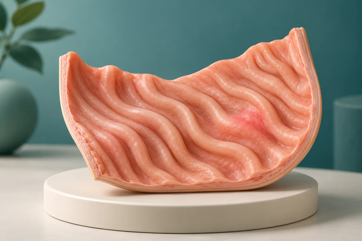
Propofol versus conscious sedation

What each option feels like, who gives it, recovery arrangements and how to discuss the choice.
Explore the guide↗
The endoscopy journeySix linked stages in a zigzag path from referral, booking and preparation to the procedure, recovery and results. Select a stage in the controls to read what happens.01Referral02Booking03Preparation04Procedure day05Recovery06ResultsYour steps and timing depend on your needs and the service arranging the procedure.
Procedures and follow-up

Your endoscopy: from referral to results

A clear guide to how an endoscopy is arranged, what happens on the day, and when you can expect results.
Explore the guide↗
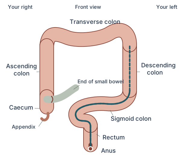
Whole colon with the lower-bowel examination route
Procedures and follow-up

Flexible sigmoidoscopy

A shorter examination of the lower bowel: when it helps, enema preparation, sedation and recovery.

Explore the guide↗
Front view of the stomach, food pipe and duodenumThe food pipe enters the upper stomach. The stomach lies mainly on the patient’s left, on image right. Its outlet, the pylorus, leads to the duodenal bulb and a C-shaped duodenum. Routine gastroscopy examines the first two parts of the duodenum, not the whole small bowel.
Procedures and follow-up

Gastroscopy: what to expect

Where the camera goes, biopsies, preparation, sedation and your results.

Explore the guide↗
Front view of the large bowel and colonoscopy routeThe ascending colon and caecum are on the patient’s right, on image left. The transverse colon crosses the upper abdomen, the descending colon is on the patient’s left, and the sigmoid joins the rectum. The terminal ileum enters the inner side of the caecum. The appendix branches from the caecum and is not part of the camera route.
Procedures and follow-up

Colonoscopy: what to expect

How the bowel is examined, polyp removal and planning your preparation and recovery.

Explore the guide↗
A negative FIT is one part of the assessmentA stool sample is checked for blood. A negative FIT is followed by assessment of symptoms and history to decide whether further tests are needed. This is an explanatory flow, not a personal risk calculator.FIT sampleChecks for bloodNegative FITDoes not excludeevery causeSymptomsHistoryNext testsThe next step depends on the whole clinical assessment.
FIT and bowel testing

My FIT was negative, but my bowel symptoms continue

A negative FIT makes bowel cancer less likely, but it does not explain your symptoms. If bleeding, a change in bowel habit or pain continues, arrange a review with your GP or the team looking after you. Do not wait for your next screening kit if symptoms persist or get worse.

Explore the guide↗
Usual relationship of the food pipe, diaphragm and stomachFront-view schematic. The stomach lies mainly on your left, on the right of this picture. The junction is below the diaphragm. Only the food pipe, diaphragm and upper stomach are shown. Their exact shape and position vary.ChestAbdomenYour rightYour leftDiaphragmFood pipeStomachJunction
Common conditions

Hiatus hernia: what does this finding mean?

A hiatus hernia means part of the stomach sits above the diaphragm. It is a common finding at gastroscopy and often needs no treatment. I check the type of hernia, your symptoms and the other findings before recommending what to do.

Explore the guide↗
Diverticulosis commonly affects the sigmoid colonA simplified front-view colon is shown with several small pouches attached to the sigmoid segment. The pouches are shown as an example of diverticulosis, not as a personal test result.ColonSigmoid colon — common site
Common conditions

Diverticulosis and diverticulitis: what is the difference?

Diverticulosis means there are small pouches in the wall of the colon. They are common and usually cause no symptoms. Diverticulitis is different: a pouch and the tissue around it have become inflamed. A scan or colonoscopy report may mention diverticulosis even when it is not causing your symptoms.

Explore the guide↗
Biopsy from the surface liningThe biopsy forceps enter from the lumen and grip a small part of the mucosa. The submucosa and deeper muscle are not cut through in this routine-biopsy illustration.Inside the digestive tractSurface lining (mucosa)SubmucosaDeeper muscleBiopsy forceps
Procedures and follow-up

Biopsies after endoscopy: why were samples taken?

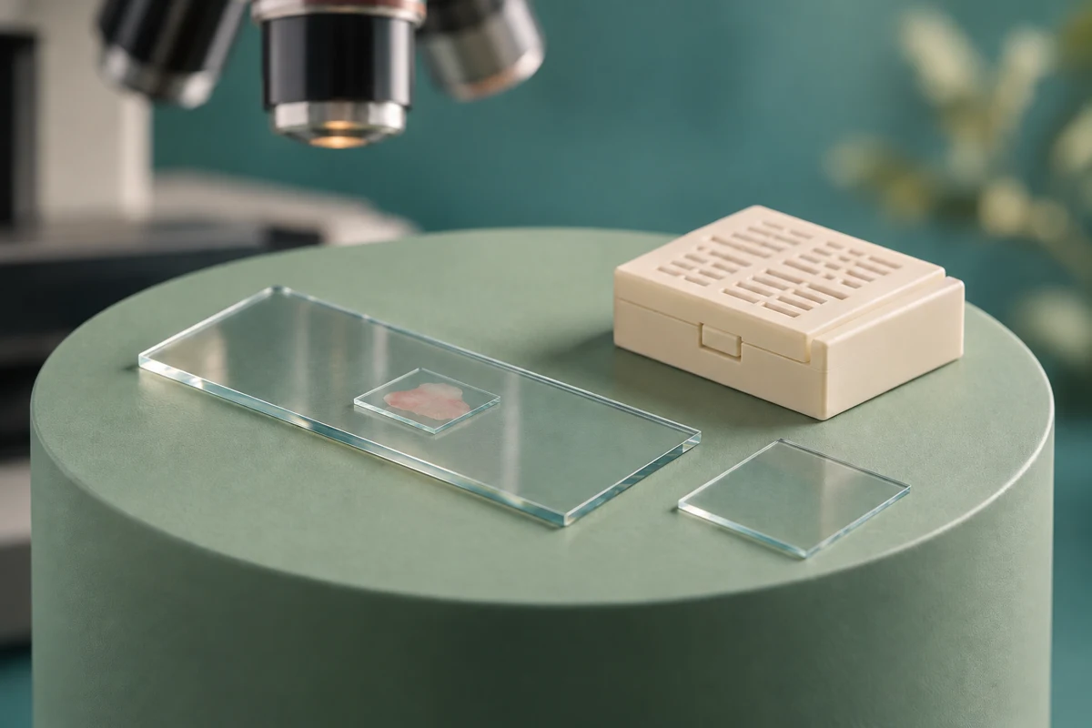
A biopsy is a small tissue sample sent to a laboratory. Taking one is routine during many endoscopies and does not mean I have found cancer. A pathologist can see changes under the microscope that are too small to see with the camera.

Explore the guide↗
Usual relationship of the food pipe, diaphragm and stomachFront-view schematic. The stomach lies mainly on your left, on the right of this picture. The junction is below the diaphragm. Only the food pipe, diaphragm and upper stomach are shown. Their exact shape and position vary.ChestAbdomenYour rightYour leftDiaphragmFood pipeStomachJunction
Common conditions

Reflux and oesophagitis: symptoms, findings and treatment

Reflux happens when stomach contents come back up into the food pipe. It can cause heartburn or an acid taste. Oesophagitis means the lining is inflamed. The two often occur together. I use your symptoms and test results to recommend treatment.
Explore the guide↗
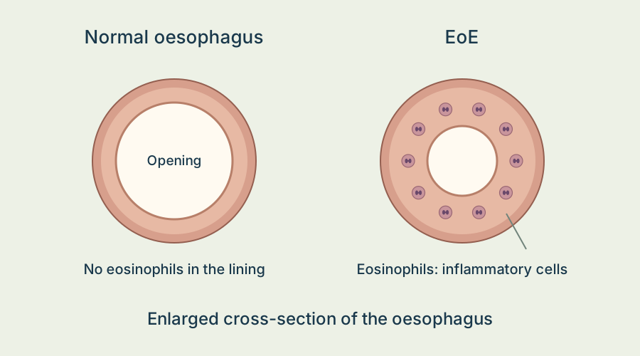
Normal oesophagus compared with inflammation in EoE
Common conditions

Eosinophilic oesophagitis (EoE)

Food sticking and difficulty swallowing: diagnosis, medicines, diet, dilatation and follow-up, with two short films.

Explore the guide↗
Fat within liver cellsThe liver is shown on the left of the diagram beside an enlarged simplified tissue concept. Yellow circles represent fat droplets within liver cells. Red dots in the inflammation view represent inflammatory cells. Purple bands in the fibrosis view represent scar tissue between cells. These are separate examples, not stages that everyone passes through or a real histology image.LiverSimplified tissue concept
Liver, gallbladder and pancreas

Fatty liver: what does my scan mean?

A scan may show fat in your liver. That result alone cannot tell whether there is inflammation or scarring. I look for possible causes and check whether you might have significant scarring, because that affects whether you need more tests or follow-up.

Explore the guide↗
Where a gallstone sits changes the problemSimplified front-view cutaway. The liver is mainly on your right, at image left. The gallbladder is underneath it. Its cystic duct joins the common hepatic duct to form the common bile duct, which drains through the medial wall of the descending duodenum. The pancreas, pancreatic duct, stomach and many smaller branches are omitted. The yellow stone marks one illustrative location; this is not an image of your own anatomy.Your rightYour leftLiverGallbladderCystic ductCommon bile ductDuct openingDuodenumFront-view cutaway. Other organs and some duct branches are omitted.
Liver, gallbladder and pancreas

Gallstones: a scan finding or the cause of pain?

Gallstones are often found on a scan done for another reason, and many cause no symptoms. I check whether the pain fits gallstone pain, where the stones are and whether there is a blockage or inflammation. Finding a stone does not by itself mean you need an operation.

Explore the guide↗
Prepare for endoscopy: instructionsFour checklist items: location and arrival, food and clear fluids, bowel preparation if needed, and contacting our team with questions.Your personal preparation checklistCheck your location and arrival timeStop solid food before the test; clear fluids until cut-offIf having colonoscopy, collect the prescribed bowel preparationContact our team if anything is unclearYour exact instructions will be communicated directly.
Procedures and follow-up

Preparing for endoscopy: a practical checklist

Good preparation helps me examine the lining clearly and carry out your procedure safely. Your exact food, fluid, medicine and bowel-preparation instructions will be communicated to you directly. Read them early so we have time to resolve any questions.

Explore the guide↗
Understanding histology: siteThe selected view explains site. Specimens are labelled by location.Where did the sample come from?OesophagusStomachDuodenumColonFood-pipe liningGastric liningSmall-bowel liningLarge-bowel liningThe location changes how a tissue finding is interpreted.
Procedures and follow-up

Understanding your histology report

A histology report describes tissue samples taken during endoscopy, including biopsies or removed polyps. Some of its words are hard to interpret without the endoscopy report. I read both reports together, then explain what the finding means for your treatment and follow-up.

Explore the guide↗
What was removed?Number: How many polyps were found?; Size: How large were they?; Type: What did histology show?; Cell changes: Was dysplasia present?. These findings help guide surveillance after removal.What was removed?NumberHow many polyps were found?SizeHow large were they?TypeWhat did histology show?Cell changesWas dysplasia present?These findings help guide surveillance after removal.
Procedures and follow-up

Follow-up after bowel polyp removal

The timing of your next colonoscopy depends on what was found, whether the polyp was fully removed and how clearly I could see the bowel. I also consider the tissue result and any previous polyps. Your endoscopy report and/or histology report will record the recommended follow-up once tissue results are available.

Explore the guide↗
Usual lining where the food pipe meets the stomachFront-view cutaway, not to scale. The food pipe joins the stomach. The usual food-pipe lining ends at the join with the stomach. No Barrett’s segment is drawn in this view. The usual example and Barrett’s example keep the same outer shape and junction position. The stomach outlet, diaphragm and most of the digestive tract are omitted.Your rightYour leftUsual food-pipe liningFood-pipe / stomach joinFood pipeStomachSimplified cutaway. Only part of the upper tract is shown.
Endoscopy findings

Barrett's oesophagus: what does the finding mean?

Barrett's oesophagus is a change in the lining of the lower food pipe that can develop as a complication of long-standing reflux. It is not cancer, and most people with Barrett's do not develop cancer. I review the gastroscopy and biopsy findings together to recommend reflux treatment and any further checks.

Explore the guide↗
The pancreas and its drainage into the duodenumFront-view simplified schematic, not to scale. Your right is image left. The head of the pancreas lies in the curve of the duodenum on your right. The body and tail extend to your left, mainly behind the stomach, whose outline is dotted. The pancreatic duct runs through the gland towards the head and opens into the descending duodenum. In this common arrangement it joins the bile duct near the opening. Small branches, the liver, gallbladder, spleen and blood vessels are omitted. In the EUS view, a partial probe symbol sits inside the stomach outline; a shaded conceptual ultrasound fan points towards the pancreas.Your rightYour leftStomach outlineBile ductDuodenumHeadBodyTailPancreatic ductOpening into the bowelFront view. The pancreas is mainly behind the stomach.
Liver, gallbladder and pancreas

Pancreatic cancer: symptoms, tests and who needs screening

If you are worried about pancreatic cancer, tell me what prompted the concern: a symptom, a scan result or a family history. Each of these situations needs a different approach. Regular screening is usually considered only for people with a high inherited risk, not for everyone.

Explore the guide↗
The small bowel connects the stomach to the large bowelSimplified front view, not to scale. Your right is image left. The food pipe enters the stomach, which lies mainly on your left. The stomach outlet leads into a C-shaped duodenum on your right. Its distal portion crosses the midline and rises towards the duodenojejunal flexure on your left before continuing into coils of jejunum and ileum. The terminal ileum joins the inner, medial side of the caecum through the ileocaecal valve. Only the caecum, a short ascending-colon segment and the appendix are shown from the large bowel. The rest of the colon, other organs, mesentery and blood vessels are omitted. There is no drawn boundary between jejunum and ileum because their change is gradual and their loops do not have fixed positions. Your rightYour leftFront view Food pipe Stomach Duodenum Stomach outlet Jejunumand ileum Caecum End of the ileum joins the caecum Only the beginning of the large bowel is shown. Bowel loops vary. Their shape and relative lengths are simplified.
Procedures and follow-up

Looking at the small bowel: capsule cameras, scans and enteroscopy

Gastroscopy and colonoscopy do not examine most of the small bowel. I recommend another test when your symptoms or earlier results give us a reason to look there. I choose the test based on what we need to find, such as bleeding, inflammation, a narrowing or an area that needs a biopsy.

Explore the guide↗
Different tests answer different questionsThe sequence is planned while gluten is still in the diet. Blood antibodies An immune response signal Interpret with total IgA. Duodenal biopsy Microscopic tissue assessment Usually through gastroscopy. HLA gene test Can help exclude in selected cases A positive gene is not a diagnosis. Specialist review Results, gluten intake and history Agree the confirmation pathway. Do not start a gluten-free diet just to see whether a test improves.Different tests answer different questionsThe sequence is planned while gluten is still in the diet.Blood antibodiesAn immune response signalInterpret with total IgADuodenal biopsyMicroscopic tissue assessmentUsually through gastroscopyHLA gene testCan help exclude in selected casesA positive gene is not a diagnosisSpecialist reviewResults, gluten intake and historyAgree the confirmation pathwayDo not start a gluten-free diet just to see whether a test improves.
Common conditions

Coeliac disease: getting the diagnosis right, then staying well

Coeliac disease is an immune reaction to gluten that can damage the small bowel. The treatment is a lifelong gluten-free diet, so I want to confirm the diagnosis before you change what you eat. Stopping gluten first can make the blood tests and biopsies harder to interpret.

Explore the guide↗
H. pylori beside the stomach liningSide-view microscopic concept, not whole stomach anatomy. The stomach lumen is above a protective mucus layer. Spiral symbols in the mucus represent H. pylori near the epithelial surface. The surface dips into gastric pits. Deeper glands, individual cells and muscle layers are omitted. No organisms are drawn invading the deeper wall. The scale is exaggerated; this is not a clinical photograph or a prediction of cancer.H. pylori lives beside the stomach lining Inside the stomachAcid-containing space Mucus layerIllustrated bacteria Surface lining and gastric pits, simplified Most infected people do not have an ulcer or cancer.
Common conditions

H. pylori: testing, treatment and checking it has cleared

Helicobacter pylori (H. pylori) is a bacterium that can live in the stomach lining. A reliable positive test for active infection does not mean cancer. I check which test you had and which treatment suits you. After treatment, you need another test to confirm the infection has cleared, even if you feel better.

Explore the guide↗
Where faecal calprotectin comes fromSimplified side view of bowel tissue. White circles with lobed centres represent neutrophils, a type of inflammatory white blood cell. A migration arrow points from tissue towards the bowel lumen, with dots representing calprotectin released into bowel contents. A stool container represents laboratory sampling. This is not a quantitative model, histology slide or complete immune pathway. Tissue and sample are separate scales. No diagnostic threshold is shown.A signal of inflammation in a stool sample Calprotectin comes largely from inflammatory white blood cells. Inside the bowelTissue below the surface Calprotectin Stoolsample The result cannot identify the cause of inflammation by itself.
FIT and bowel testing

Faecal calprotectin: what does an inflammation test in poo tell us?

Faecal calprotectin is a poo test that can show whether there is inflammation in the bowel. It does not diagnose Crohn's disease, ulcerative colitis or cancer. I read the result with your symptoms, medicines and other test results before recommending what to do next.
Explore the guide↗
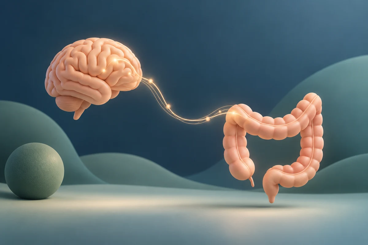
Conceptual illustration of communication between the gut and brain
Common conditions

IBS: understanding your symptoms and choosing treatment

Irritable bowel syndrome (IBS) can cause recurring abdominal pain, bloating, constipation or diarrhoea. It can disrupt meals, sleep, work and travel. I ask what your symptoms are like, check for warning signs and use a small number of tests when they are needed. Then we can agree on treatments to try and when to review them.
Explore the guide↗
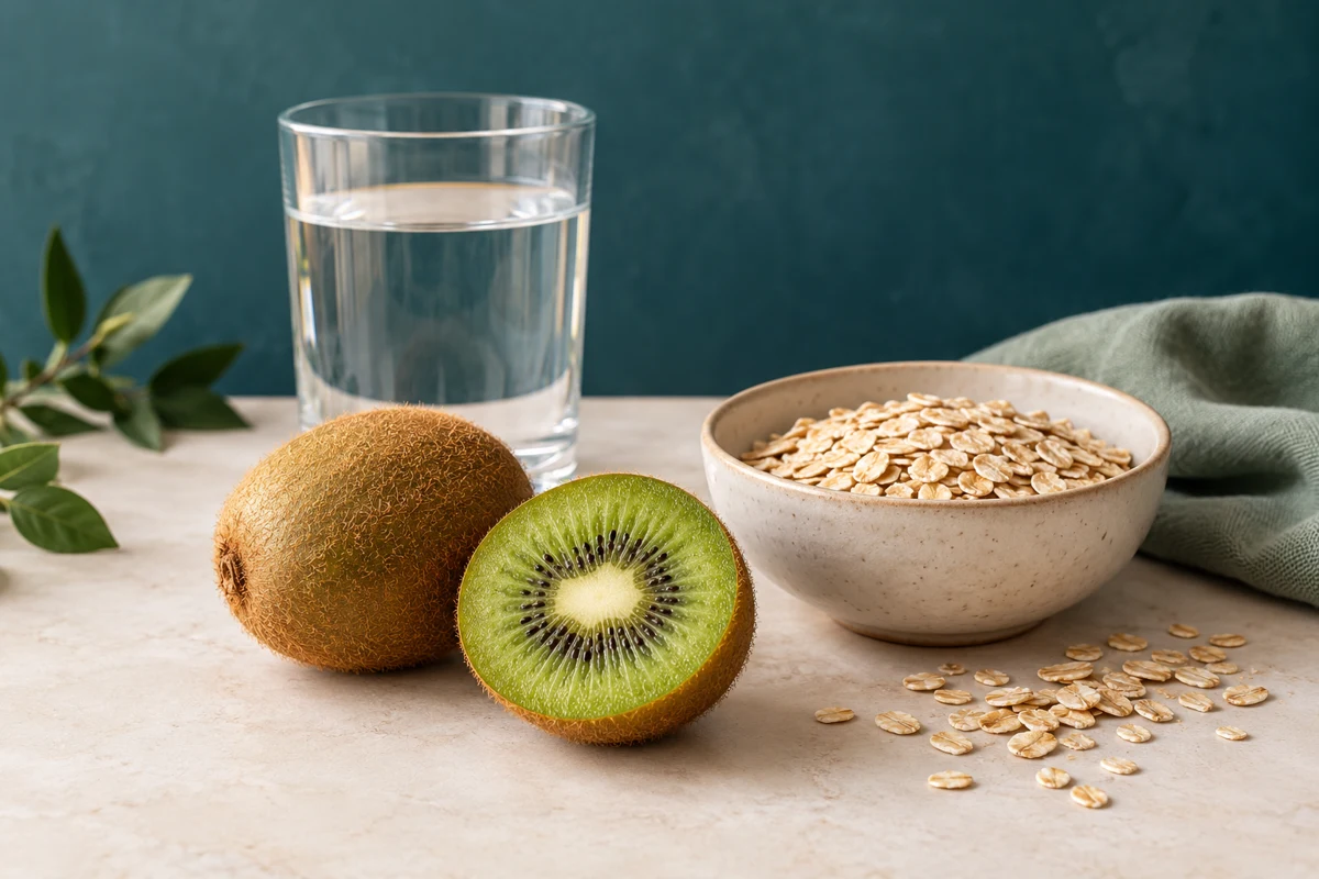
Kiwifruit, water and oats, as examples of food and fluid choices
Common conditions

Constipation: softer poo and easier emptying

Constipation can mean hard poo, straining, going less often or still feeling blocked after you go. You can be constipated even if you have a bowel motion every day. I ask about your poo, emptying, medicines and other health conditions before choosing treatment.

Explore the guide↗
Topics in preparation
  • Iron deficiency: why further investigation may be needed
  • Bile acid diarrhoea
  • Understanding abnormal liver blood tests

Arrange an appointment

Assessment of digestive symptoms, endoscopy and advice on further investigations. Auckland appointments and telehealth.

Contact our team